Radiologists, pediatricians, orthopedists, emergency room physicians, and rehabilitation physicians will make use of this quick-access visual reference to determine, for example, whether a particular bone configuration is normal for a child or an indication of problems. Roughly 2,300 images present views of the skeleton at every developmental milestone. Organization is by gender and body part. The included DVD provides electronic access to the images. The four authors are affiliated as follows: S. Lowell Kahn (Tufts U.), Cree M. Gaskin (U. of Virginia Health System), Victoria L. Sharp (Botsford Hospital, Michigan), and Theodore E. Keats (deceased; formerly with U. of Virginia Health System). Annotation ©2012 Book News, Inc., Portland, OR (booknews.com)
Bone age assessment, a crucial part of the diagnosis and management of pediatric growth disorders as well as the timing of certain pediatric orthopedic procedures, has for decades depended on the meticulous examination of plain radiographs. Examining the subtle changes present within the maturing human hand often proves to be challenging and time-consuming.
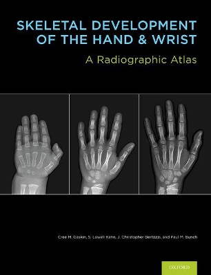
Building on the popular Greulich and Pyle atlas, this book modernizes the method for pediatric skeletal maturity determination. It offers a wealth of images, carefully mined from thousands of digital radiographs from University of Virginia's Picture Archiving and Communication System (PACS), edited to best demonstrate important developmental bone features, and organized by age and sex for rapid reference. To expedite learning and clinical image analysis, images come in pairs: annotated and unannotated, for easy comparison. Succinct annotations on the images replace lengthy text to provide a quicker and clearer understanding of the skeletal age. These annotations highlight important and subtle features to help distinguish images that otherwise look superficially alike. The result is an atlas of exceptionally high quality skeletal radiographic standards that capture both the major and finer details of the accepted standards of Greulich and Pyle.
The user-friendly format of this book enables a faster, more accurate, and more educational approach to determining skeletal maturity. The Digital Bone Age Companion packaged with the book is a computer program that facilitates viewing of the atlas images in digital format. Users can easily zoom in on radiographic features, set image level and width to their preference, and compare two or three reference standards side-by-side for difficult cases. Most importantly, the program expedites evaluation, optimizes workflow, and minimizes user-introduced errors with the reliable bone age calculator and built-in report generator. The digital format may also be available for integration with your Radiology Information System (RIS) for further workflow enhancement.
Given the broad application of pediatric bone aging, Skeletal Development of the Hand and Wrist is not only intended for practicing and training radiologists, but for all of those who employ bone age studies as part of their practice.


 Daugiau apie elektronines knygas